
ここからコンテンツです。
How Capturing the Movement of Ions can Contribute to Brain Science and Improve Disease Diagnosis
Kazuaki Sawada
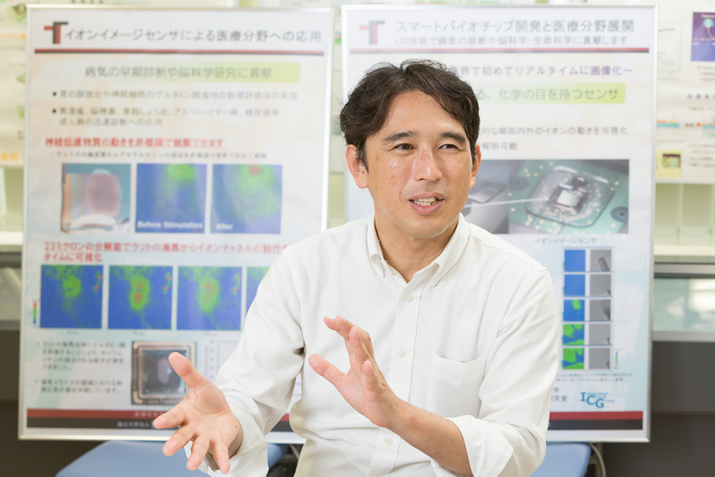
Professor Kazuaki Sawada’s work focuses on the development of biosensors that combine integrated circuit technology and sensor technology. Currently Prof. Sawada’s main project is working on a "bio image sensor", which he hopes to use to directly observe ion distribution and movement as visual images, in order to contribute to the development of brain science and disease diagnosis. There is growing excitement in the scientific community regarding the potential practical applications of this innovative sensor, which Prof. Sawada is striving to fulfill by establishing research associations and incorporated associations through industry-university.
Interview and report by Madoka Tainaka
Development of world's first sensor that can detect ion reactions
Today, the major types of image sensors for acquiring two-dimensional images by sensing are CCD (Charge-Coupled Device) and CMOS (Complementary metal-oxide-semiconductor). These semiconductor devices are mounted on digital cameras and smartphone cameras. They capture the amount of light as an amount of electric charge and process it after converting it to an electric signal and are widely used not only for cameras but also for measurement of magnetic fields and voltages. However, until now, there were no sensors that could directly capture the distribution and movement of ions, in the way that Prof. Sawada’s bio image sensor is now able to do.
"As a means for measuring ions, using litmus paper for measuring hydrogen ions is well established, but what sets our device apart is that it can measure the amount. This is the world’s first sensor that can directly observe the movement of ions," says Prof. Sawada.
The trigger for development dates back to Prof. Sawada’s fourth undergraduate year when he was a student at Toyohashi University of Technology. In response to a request from a company, he worked on the development of a hydrogen ion sensor through an industry-university collaboration that became his graduation thesis. However, the sensor didn’t achieve the desired performance and the research ended there. After that, Prof. Sawada worked for a time on material development, but after doing some research on image sensors at Shizuoka University more than ten years later, he decided to once again take on the challenge of becoming the first person to successfully image ion changes.
"As a mechanism, we read the movement of ions from the change in potential of a semiconductor (ISFET: Ion Sensitive Field Effect Transistor) by means of a sensitive membrane of ions placed on the surface of the semiconductor. It’s a little complicated, but at that time, depending on the concentration of hydrogen ions, a state called a "potential well" in quantum theory is formed on the semiconductor surface. To put it simply, the depth of the cup changes according to the concentration of ions. To measure the depth of the cup, we inject a lot of electric charge into it and measure the amount of charge that accumulates. We can then measure the electric charge by converting it into a voltage like a CCD or CMOS image sensor. In this way, we can read the ion concentration with a high level of sensitivity," explains Prof. Sawada.
In 1997, Prof. Sawada acquired a patent for this innovative method, and since then has continually strived to further develop and improve the bio image sensor.
As the accuracy improved we became aware of the potential goals
Unfortunately it was not possible to achieve the target accuracy straight away. "At the beginning of the development, the number of pixels was just 1×8, and by 2005 we achieved 10×10, but at this level we could just about sense the change in ions when we dropped orange juice on the sample. At this level, nobody was really interested. However, at a scientific meeting in 2007, when we increased the number of pixels to 32×32 and used the sensor to show rice roots emitting citric acid, everyone got excited. There was a theory that plant roots emitted citric acid and dissolved nutrients in the soil to absorb them, but until then no one had been able to observe the phenomenon. After that breakthrough, researchers in various fields came to me saying they wanted to use the sensor."
Originally, the research started from the pure interest of Prof. Sawada, who wanted to try to simply observe the movement of ions. But based on people’s reactions, he began to understand how the research would be useful for society. "Doctors at the medical department told me that the ability to see ion reactions could be revolutionary in medicine. I was surprised when people told me it was so groundbreaking. So, when I brought them a prototype that I had managed to make, they were unsatisfied, explaining that they needed at least 100 pieces. If we do not raise the accuracy and usability to a level that has the impact of making potential users want to use the technology, it will not become widely used. This revelation changed the philosophy of my research. Since then, my goal has been to fulfill my responsibility to contribute to society, whether or not the research can be published as a paper."
After that, in 2011, Prof. Sawada succeeded in observing the release of acetylcholine from a rat cerebral cortex without labels with a 128×128 (16,000 pixels) element. Furthermore, in 2014, he observed the release of potassium ions by stimulating a rat hippocampus with glutamic acid.
"Usually, a wet sample will break as soon as you place it on a chip. That was the tricky part, which explains why it took time to develop the initial idea. However, by enhancing the waterproofing of the chips, we made it possible to capture the appearance of ions leaking out of a cell, which no one had ever seen before. With these unprecedented tools that enable us to look at cells from the outside, we anticipate the potential for creating new academic fields."

In addition to that, Prof. Sawada is also working on research such as the development of a sensor that separates excitation light and fluorescent light. The key point is that it does so without using the kind of expensive optical filter that is usually indispensable for fluorescence microscopy. What is more, it can visualize the fluorescence of samples with the same accuracy as a fluorescence microscope. As a result of such discoveries, Prof. Sawada explains that he receives endless daily inquiries from companies and research institutions, especially from the medical field.
Promoting social implementation through two pillars: the study group and the council
At present, the number of pixels of the element of the bio image sensor has reached 1.3 million (1280×960). This has been achieved through industry-university collaborative development. In order to advance implementation in society, in 2012, Prof. Sawada established the "Multimodal Bio Image Sensor Study Group", of which he is the chairman. About 25 organizations including companies such as semiconductor manufacturers, universities, and national research institutions have participated and are deepening consideration of the industrial contribution of multimodal bio image sensors and the creation of new businesses.
Meanwhile, in September 2016, Prof. Sawada founded a general incorporated association named "Toyohashi Sensor Council". The council is accelerating initiatives to create bio sensor markets, such as determining standards and specifications, holding technical lectures and providing consultancy.
"Since our research group is after all a university organization, there were aspects that were difficult to control from the perspective of fairness, such as how to handle patents in promoting commercialization. So I decided to devote myself to research and transferred the license of approximately 70 patents owned by the university to the council, and we made the council responsible for its development as a business," says Prof. Sawada.
Incidentally, since the press release, more and more people are asking to participate in the council, but Prof. Sawada says not everyone can join.
"Since our goal is not to make money, but to return the intellectual property of the university to society and to maximize the social utility of the sensor, I select companies to participate based on their willingness to get involved on a volunteer basis. We’re expecting to be able to announce a new product next year, so hopefully that’s something we can all look forward to."
It is indeed exciting to have such innovative achievements on the horizon, especially when they are the product of a researcher’s simple passion to know the unknown, and to be of service to society.
Reporter's Note
Prof. Sawada's goal to become a researcher started from his love of Antarctica. "I wanted to join the Japanese Antarctic Research Expedition, but I didn’t feel I could become a geologist or meteorologist. However, I thought that I might be able to work in communications, so I joined the engineering department. Actually, I joined the new Department of Electrical and Electronic Information Engineering. In the end, I did not study communications at all, and my teacher advised me that sensors will become important in various fields in future, so I proceeded down the road of sensor development," says Prof. Sawada.
Although Prof. Sawada says he has "completely moved on from his early goal," he has not thrown away his dream of traveling to Antarctica. "Sensors should be indispensable for measuring ice sheets and soil. I would like to see this unknown world that nobody has seen yet. I would be delighted to go if I was asked," he laughs. In this way, Prof. Sawada shows himself to be a researcher who is driven by curiosity and who purely wants to explore the truth.
イオンの動きを捉え、病気の診断や脳科学に役立てたい
澤田和明教授が手がけるのは、集積回路技術とセンサ技術を融合させた、さまざまなバイオセンサの開発である。その代表が「バイオイメージセンサ」で、イオンの分布や動きをダイレクトに画像として見ることにより、脳科学の進展や病気の診断などに役立てたいという。従来にない画期的なセンサとして、実用化への期待が高まる中、澤田教授は産学連携で研究会や社団法人を設立、実用化に向けた動きに取り組む。
世界初、イオン反応を見るセンサを開発
現在、センシングにより二次元の画像を取得するイメージセンサといえば、デジカメやスマートフォンのカメラに搭載されている半導体素子(CCD=Charge-Coupled DeviceやCMOS=Complementary metal-oxide-semiconductor )が主流である。これは光の量を電荷の量として捉え、電気信号に換えて処理するもので、カメラのほか、磁界や電圧の計測などにも幅広く活用されている。しかしこれまで、澤田教授が手がける「イオン」の分布や動きをダイレクトに捉えるようなセンサは存在しなかった。
「イオンを測るものとしては、水素イオンを測るリトマス試験紙が有名ですが、これは量を測るもの。イオンの動きを直接目で見るセンサの開発は世界初の試みです」と澤田教授は言う。
開発のきっかけは、澤田教授が豊橋技科大の在学生だった学部4年次に遡る。企業からの要請を受け、産学連携で水素イオンセンサの開発を手がけて卒論にまとめた。しかし、思うような性能は出せず、そのまま研究は終了。その後は材料開発の研究をしていたが、十数年後、静岡大学でイメージセンサの研究をする中、まだ誰も手がけていないイオン変化のイメージングにふたたび挑戦することにした。
「仕組みとしては、半導体中にイオンの感応膜を置いた半導体(ISFET:Ion Sensitive Field Effect Transistor)の電位の変化からイオンの動きを読み取ります。少し難しい話になりますが、その際に水素イオンの濃度に応じて半導体表面に、量子論で言うところの“ポテンシャルの井戸”と呼ばれる状態が形成されるのです。噛み砕いて言えば、イオンの濃度によってコップの深さが変化するということ。そのコップの深さを測るためには、コップにたくさん電荷を注入して、そこに溜まった電荷量を計測すればいい。そうすれば、CCDやCMOSイメージセンサと同じように電荷量を電圧に変換して計測することができます。これにより、イオンの濃度を高感度に読み取ることができるのです」と澤田教授は説明する。
1997年、澤田教授はこの画期的な方式の特許を取得して、以後、バイオイメージセンサの開発に邁進していくことになる。
精度が向上するにつれ出口が見え始めた
しかし、最初から狙った精度を出せたわけではない。
「開発当初は画素数が1×8と少なく、2005年には10×10が開発できましたが、みかんの汁を落としたことによるイオン変化がなんとなくわかる程度で、誰にも見向きもされませんでした。ところが2007年にある学会で、32×32に画素数を増やした素子を使って、稲の根がクエン酸を放出している様子を映したら、会場がざわついたのです。植物の根がクエン酸を出し、土壌の養分を溶かして吸っているという説はありましたが、それまでその様子を誰も見たことがなかったからです。その後、さまざまな分野の研究者から、使ってみたいと声がかかるようになりました。」
当初は、イオンの動きを見たいという澤田教授の純粋な興味で始まった研究だったが、まわりの反応に触れるにつれ、研究がどう社会に役立つかを意識するようになっていったという。
「医学部の先生から、イオンの反応が見えれば医学の常識が変わる。それくらい画期的なことだよ、と言われて驚きました。そこで、なんとか作った試作品を持参すると、『少なくとも100個はないと使えない』と怒られてしまいました。使いたい人が、使ってもいいと思うようなインパクトのあるレベルまで精度と使い勝手を上げなければ、社会に浸透することはありません。以来、たとえ論文にはならなくても、社会との橋渡しをしていくことも、研究者の務めだと強く感じるようになりました。」
その後、2011年には、128×128(1.6万画素)の素子でラットの大脳皮質からアセチルコリンが放出される様子を非標識で観察することに成功。さらに、2014年には、ラットの海馬をグルタミン酸で刺激することでカリウムイオンが放出される様子を捉えた。 「通常、ウェットな試料をチップに載せるとすぐに壊れてしまいます。構想から開発まで時間がかかったのはそのためです。チップの耐水性を高めたことで、これまで誰も見たことのない、細胞の外側にイオンが浸み出す様子を捉えることができるようになりました。細胞を外側から見るという、これまでになかった道具ができたことで、新しい学問領域を生み出すことができるのではないかと期待しています」
そのほかにも澤田教授は、蛍光顕微鏡に欠かせない高価な光学フィルタを使用することなく、励起光と蛍光光を分離し、蛍光顕微鏡と同程度の精度で試料の蛍光を可視化するセンサの開発なども手がけている。こうした成果を背景に、医学分野を筆頭に、企業や研究機関からの引き合いが絶えない日々だという。
研究会と協議会の二本柱で社会実装を推進
現在、先のバイオイメージセンサの素子の画素数は130万(1280×960)にまで達している。その背景にあるのが、産学連携による開発だ。澤田教授は社会実装を進めるために、2012年、自らが会長を務める「マルチモーダルバイオイメージセンサ研究会」を設立。半導体メーカーなどの企業、大学、国の研究機関など約25 の組織が参画して、マルチモーダルバイオイメージセンサの産業貢献や新規事業創出に向けた検討を深めている。
一方、2016年9月に「一般社団法人 豊橋センサ協議会」を設立。規格や仕様の決定、技術講習会の開催、コンサルティングなど、バイオセンサの市場創出に向けた取り組みも加速させている。
「研究会はあくまでも大学の一組織なので、製品化を進めるうえで、特許の扱いをどうするかなど、公平性の観点からコントロールするのが難しい面がありました。そこで、私は研究に専念することにして、大学が持つ約70の特許の実施権を協議会に移し、ビジネスとしての発展は協議会が担うかたちにしました」と澤田教授。
ちなみに、報道発表以来、協議会に参加したいという申し出が増えているが、誰でも加入できるわけではないという。
「お金儲けではなく、大学の知財を社会に還元し、開発したセンサを社会に広めていくことが目的なので、実際に手を動かして、手弁当で関わってくれる人たちに参加してもらいたいという思いから、企業を選別しています。来年には、新製品の発表ができると思いますので、期待していてください。」
未知のものを知りたいという研究者の思いと、それが社会に還元されることの喜びが原動力となって、大きな実を結びつつある成果の発表が待ち遠しい。
(取材・文=田井中麻都佳)
取材後記
澤田教授が研究者を目指したきっかけは、南極への憧れだったという。「南極越冬隊員に加わりたかったのですが、地質学者や気象学者にはとてもなれそうもなく。でも、もしかしたら通信士ならできるかもしれないと思い、工学部へ進学しました。ところが、入ったのは新しくできた電気電子工学科。結局、通信の勉強は一度もすることなく、恩師から、今後、さまざまな分野で重要になるとアドバイスを受けて、センサ開発の道に進みました」と澤田教授。
「すっかり初心を忘れてしまいました」と言うものの、南極への夢は今も捨てていない。「氷床や土壌の計測にセンサは欠かせないはず。まだ誰も見たことのない未知の世界をぜひ見てみたい。声がかかれば、喜んで行きますよ」と笑う。そこには、好奇心に駆られ、純粋に真理を探究したいという研究者の姿があった。
Researcher Profile
Dr. Kazuaki Sawada
Dr. Kazuaki Sawada received his B.A. and M.S. degrees in electrical and electronic engineering in 1986, 1988, respectively, and he received a Ph.D. degree in system and information engineering in 1991, all from Toyohashi University of Technology, Aichi, Japan.
From 1991 to 1998, he was a Research Associate in the Research Institute of Electronics, Shizuoka University, Shizuoka, Japan. Since 1998, he joined the Department of Electrical and Electronic Engineering, Toyohashi University of Technology, where he is now serving as a Full Professor.
Reporter Profile
Madoka Tainaka is a freelance editor, writer and interpreter. She graduated in Law from Chuo University, Japan. She served as a chief editor of “Nature Interface” magazine, a committee for the promotion of Information and Science Technology at MEXT (Ministry of Education, Culture, Sports, Science and Technology).
ここでコンテンツ終わりです。