
Multisensory information detection using multi-channel electrocorticography film that can be placed over a wide area of the cerebral cortex
Development of electrocorticography device for simultaneous detection of multi-modality information in the mouse brain Hiroto Sekiguchi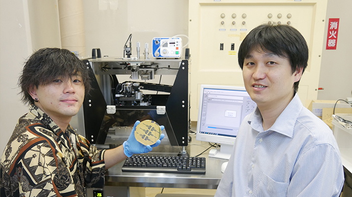
A research group led by Associate Professor Hiroto Sekiguchi from Toyohashi University's Department of Electrical and Electronic Information Engineering, along with Assistant Professor Susumu Setogawa and Associate Professor Noriaki Ohkawa from Dokkyo Medical University's Comprehensive Research Facilities for Advanced Medical Science (Associate Professor Setogawa is currently a Specially Appointed Assistant Professor at Osaka Metropolitan University), have developed a flexible electrocorticography (ECoG) (Note1) film for simultaneous detection of multisensory information (Note2) from multiple regions of the cerebral cortex by placing neural electrodes over a wide area of a mouse brain surface.
Researchers believe that the human brain achieves cognitive functions such as attention, learning, and memory through the simultaneous processing and integration of various sensory information across multiple regions of the cortex. In order to understand the neural information processing mechanisms underlying these cognitive functions, researchers required a device capable of simultaneously recording neural activity from a wide area of the cerebral cortex in rodents such as mice and rats, including the temporal region responsible for processing diverse sensory information. However, the device itself was difficult to install in the temporal region of rodents because it is obstructed by the skull and surrounding temporal muscles.
Therefore, in order to successfully position an ECoG recording device in the temporal and deep regions of the cerebral cortex of mice, it was crucial to develop a device and establish a technique which allows recording electrodes to be placed in the narrow gap between the skull and the surface of the cerebral cortex. Our research group achieved two significant advancements: (1) Development of a cortical ECoG device composed of an appropriate film capable of both the flexibility and rigidity to adhere to the brain. (2) Establishment of a surgical technique for effectively attaching the device to the temporal region of the brain. This measurement technique using a new ECoG measurement device has achieved a wider range of neural activity measurement than existing measurement techniques, and is expected to lead to the development of large-scale ECoG research to clarify the mechanisms of interaction between brain regions in the future.
The results of this research were published online in the scientific journal Molecular Brain on May 3, 2023. This research was supported by the Japan Science and Technology Agency (JST) Strategic Basic Research Promotion Project PRESTO (JPMJPR1885), the Casio Science Promotion Foundation, the Toyoaki Scholarship Foundation, the Foundation of Public Interest of Tatematsu, the Research Foundation for Opto-Science and Technology, the Takeda Science Foundation, the Naito Foundation, the Astellas Foundation for Research on Metabolic Disorders, and the Tochigi Industrial Promotion Center (the Grant-in-Aid for World-Class Technological Research and Development).
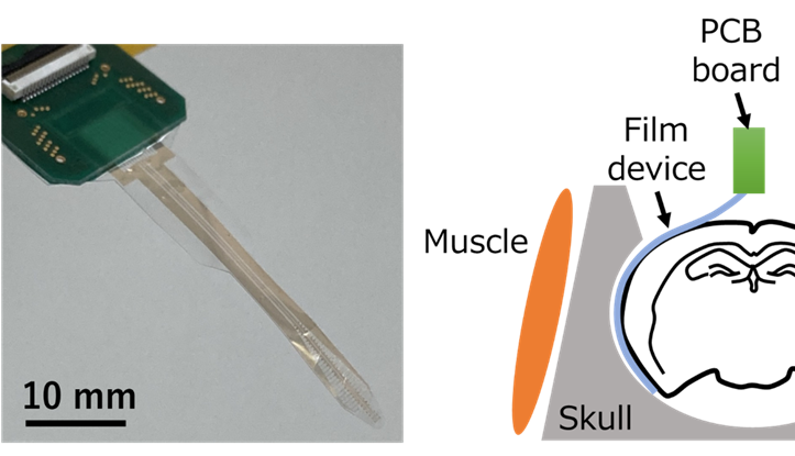
Schematic diagram of the electrocorticography method for a wide area of the brain using the developed device (right).
The total number of patients related to mental disorders such as dementia, schizophrenia, and developmental disorders is on the rise. One contributing factor to these diseases is impaired cognitive function due to the failure of information integration systems in the brain. However, the precise mechanisms by which the brain integrates diverse sensory information obtained from various environments are still not fully understood. In order to deepen our understanding of these information integration systems, it is necessary to develop biometric techniques to simultaneously monitor the activities of multiple brain regions governing diverse sensory information that are distributed across the cerebral cortex. To understand the mechanisms of the neural circuits of cognitive functions, neuroscience research has been conducted in rodents such as mice and rats, where state-of-the-art tools can be applied, and large-scale measurement techniques covering the parietal and temporal regions of the rodent cerebral cortex have been sought. However, the conventional microscope-based large-scale calcium imaging (Note3) and electrophysiological techniques (Note4) for measurement of brain activity in rodents have been limited to the parietal area of the rodent head due to the hindrance caused by the thick skull and surrounding temporal muscles. Therefore, there has been a need to develop a new device that can simultaneously detect multisensory information over a wide area of the cerebral cortex, including the temporal region, and to develop a technology to install such a device.
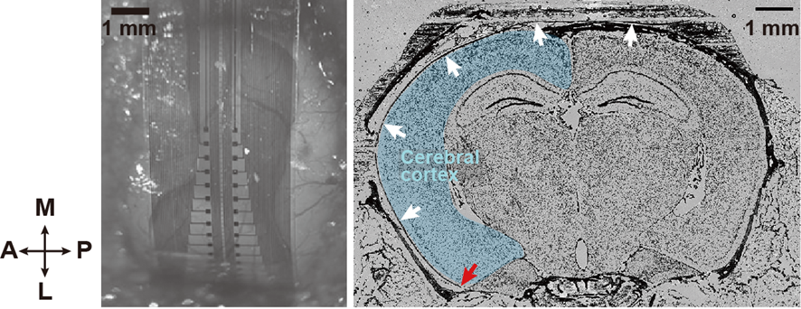
Left: Photo of the electrode array placed from the parietal region to the temporal region (device inserted into an even deeper region of the cortex).
Right: Photo of the device placed on the brain and brain surface
(white arrow = device, red arrow = device tip).
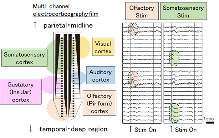
The research group aimed to realize a multi-channel ECoG measurement device that could be placed over a wide area of the brain, and conceived the idea of inserting the device into the narrow gap between the mouse skull and the dura mater. In order to insert the device into the narrow site and place it on the brain surface, it was necessary to maintain both flexible and rigid properties that would allow it to adhere closely to the brain. The research group successfully addressed this challenge by selecting the appropriate width and thickness of parylene film (Note5), which serves as the base of the device, and by establishing a surgical technique for placing the film in the temporal region. As a result, the research group was able to successfully position multi-channel electrodes across a wide area extending from the somatosensory cortex to the deepest part of the cerebral cortex, known as the olfactory cortex. Furthermore, by using an ECoG device equipped with 64-channel recording electrodes, the group successfully obtained ECoG measurement from a wide area of the cerebral cortex of awake and anesthetized mice. The research group also confirmed that the developed device, along with its placement technique, can detect neural responses evoked by both somatosensory and olfactory stimuli in the same mouse, thereby enabling simultaneous detection of widely distributed multisensory information in the brain.
The multi-channel ECoG device technology developed in this study, which can be applied to a wide area of the mouse brain, is a new measurement technology that expands the scope of ECoG recording in the cerebral cortex of rodents. Until now, ECoG measurement from the cortical surface of the temporal region was only possible with humans and monkeys. However, this innovative technology will enable electrocorticogram measurement even in mice across a wide range from the parietal region to the temporal region. There are a variety of animal models of pathological conditions in rodents that have been created using genetic engineering technology, and the newly developed measurement technology will make it possible to compare human cases with the results of research on animal models. Further understanding of the mechanisms of the brain system that integrates diverse information in rodents is expected to lead to the elucidation of pathological mechanisms of human neurological and psychiatric diseases and the development of new therapeutic techniques.
Explanation of Terms
Note1: Electrocorticography
A method of measuring neural activity in which part of the skull is surgically removed and a sheet with multiple electrodes is placed on the surface of the brain in order to electrically measure the activity of the cerebral cortex.
Note 2: Multisensory information
Refers to multiple senses such as somatosensory (touch), visual, olfactory, auditory, and gustatory. The human brain perceives phenomena by combining these multiple senses.
Note 3: Calcium imaging (method)
A method of obtaining spatiotemporal information of neural activity as image data by converting neural cell activity into fluorescence using dyes and proteins that emit fluorescence when bound to calcium ions.
Note 4: Electrophysiological technique
A method of recording electrical information (potential/current) of cells by placing glass or metal electrodes on neural cells. Examples include electroencephalograms, electrocardiograms, and electromyograms.
Note 5: Parylene film
A general term for paraxylylene-based polymers. Known as a biocompatible material. An extremely thin film can be formed using vapor deposition. Parylene film can be used as a coating material for biomedical devices such as pacemakers.
Reference
Susumu Setogawa, Ryota Kanda, Shuto Tada, Takuya Hikima, Yoshito Saitoh, Mikiko Ishikawa, Satoshi Nakada, Fumiko Seki, Keigo Hikishima, Hideyuki Matsumoto, Kenji Mizuseki, Osamu Fukayama, Makoto Osanai, Hiroto Sekiguchi, Noriaki Ohkawa, "A novel micro-ECoG recording method for recording multisensory neural activity from the parietal to temporal cortices in mice", Molecular Brain, 16, Article number:38(2023).
https://doi.org/10.1186/s13041-023-01019-9
脳広域に設置可能な多点脳波計測フィルムで多感覚情報を検出
マウス脳のマルチモダリティ情報を同時検出できる皮質脳波計測デバイスを開発 関口 寛人豊橋技術科学大学 電気・電子情報工学系 関口寛人准教授と獨協医科大学 先端医科学統合研究施設 瀬戸川将助教(現・大阪公立大学 特任助教)、大川宜昭准教授らは、マウスの脳表面の広範囲に神経電極を配置し、大脳皮質広域の複数の領域から多感覚情報(注1)を同時検出できるフレキシブルな皮質脳波計測(注2)フィルムを開発しました。
我々の脳では、複数の大脳皮質の領域が多様な感覚情報を同時に処理して統合することによって、注意、学習、記憶等の認知機能を達成していると考えられています。このような認知機能の神経情報処理の仕組みの理解に向けて、マウスやラット等のげっ歯類を対象とした多様な感覚情報を司る側頭部を含む脳の広範囲から脳波を同時検出できるデバイスが求められていました。しかし、げっ歯類の側頭部は頭蓋骨とその周囲の側頭筋に妨げられるため、デバイスを設置すること自体が困難でした。
マウスの大脳皮質側頭部・深部へ脳波記録デバイスの設置を実現するためには、頭蓋骨と大脳皮質表面の狭い隙間に記録電極を設置できるデバイス開発と手技の確立が必要でした。今回、本研究グループは、(1)脳に密着できる柔軟性と剛性の両方の性質を維持する適切なフィルムでできた皮質脳波計測デバイスの開発と、(2)脳側頭部へとデバイスを挿入する手術手技の確立によって、体性感覚と嗅覚への刺激によって誘発される大脳皮質の脳活動の検出に成功しました。新たな皮質脳波計測デバイスを用いたこの計測手法は、既存の計測技術よりも広範囲の神経活動計測を実現したことから、今後、脳領域間の相互作用のメカニズムを明らかにする大規模な皮質脳波研究の発展が期待されます。
本研究成果は、2023年5月3日に科学誌「Molecular Brain」にオンライン掲載されました。また、本研究は、科学技術振興機構(JST)戦略的創造研究推進事業 さきがけ「生命機能メカニズム解明のための光操作技術」研究領域 研究課題名「生体光刺激のための侵襲型LEDデバイスの革新」(JPMJPR1885)、カシオ科学振興財団、豊秋奨学会、立松財団、光科学技術研究振興財団、武田科学振興財団、内藤記念科学振興財団、アステラス病態代謝研究会、栃木県産業振興センター「世界一を目指す研究開発助成事業」からの支援により行われました。
日本では、認知症や統合失調症、発達障害等の精神疾患に関する総患者数は増加傾向にあります。これらの疾患の要因の1つとして、脳内での情報統合システムの障害による認知機能の破綻が挙げられていますが、ヒトや動物が多様な環境から多様な感覚情報を知覚したあと、脳内でどのように情報の統合がなされるのか、その仕組みは十分には明らかになっていません。脳がこれらの情報を統合するシステムの理解を深めるには、大脳皮質に広がって分布する多様な感覚情報を司る複数の領野の活動を同時に捉えるための生体計測技術の開発が必要です。このような認知機能の神経回路の仕組みの理解に向けて、最先端ツールが適用可能なマウスやラット等のげっ歯類を対象とした神経科学研究が進められており、げっ歯類の大脳皮質の頭頂部から側頭部をカバーする大規模な計測技術が求められていました。しかし、げっ歯類での従来の顕微鏡を用いた大規模なカルシウムイメージング(注3)や電気生理学的手法(注4)による脳波計測手法は、厚い頭蓋骨とその周囲を取り囲む側頭筋が邪魔となり、計測範囲が頭頂部に限定されていました。そのため、側頭部を含む大脳皮質の広範囲の多感覚情報を同時検出できる新たなデバイスの開発とその設置技術が求められていました。
本研究グループは、脳広範囲に設置可能な多点の皮質脳波計測デバイスの実現を目指し、マウスの頭蓋骨と硬膜の狭い隙間に滑らせて挿入することで、脳波計測デバイスを大脳皮質上に設置することを発案しました。デバイスを狭い部位へと挿入して脳表に設置するためには、脳に密着できる柔軟性と剛性の両方の性質を維持する必要がありました。目的の条件を満たすように、デバイスの基盤となるパリレンフィルム(注5)の幅と厚さを適切に選び、側頭部へと設置するための手術手技を確立することで、体性感覚野から大脳皮質の最も深部に位置する嗅覚野までの広範囲に多点電極を設置することに成功しました。また、64チャネルの記録電極を搭載した皮質脳波計測デバイスを用いて、覚醒下及び麻酔下におけるマウスの大脳皮質広域からの脳波計測を実現しました。さらに、開発した本デバイスとその設置手技によって、同一のマウスの体性感覚と嗅覚の刺激によって誘発される神経応答を検出できることを確認し、脳の広範囲に分布する多感覚情報を同時検出できることを実証しました。
本研究で開発されたマウスの脳の広範囲に適用可能な多点皮質脳波計測デバイス技術は、げっ歯類の大脳皮質における脳波計測領域をこれまでより拡大する新たな計測技術です。これまで側頭部の皮質表面からの脳波計測はヒトやサルに限定されていましたが、今回の技術によってマウスでも頭頂部から側頭部までの広範囲から脳波計測が可能になります。げっ歯類には遺伝子工学技術によって作出された多様な病態モデル動物が存在しており、今回開発した計測技術を活用することで、ヒトの症例と病態モデル動物の研究結果とを照らし合わせることが可能になります。今後、げっ歯類において多様な情報を統合する脳内システムの仕組みの理解が進めば、ヒトの神経疾患や精神疾患などの病態メカニズムの解明や、新たな治療技術の開発につながることが期待されます。
用語解説
注1:多感覚情報
体性感覚(触覚)、視覚、嗅覚、聴覚、味覚といった複数の感覚のことである。これらの複数の感覚を脳内で組み合わせることで様々なものを認知することができる。
注2:皮質脳波計測
脳活動計測の手法の1つで、外科的処置によって頭蓋骨を外し、脳表に複数の電極が並べられたシートを配置することにより大脳皮質の活動を電気的に計測する手法である。
注3:カルシウムイメージング(法)
カルシウムイオンと結合して蛍光を発する色素やタンパク質を用いて神経細胞の活動を蛍光に変換することで、画像情報として神経活動の時空間的情報を得る手法である。
注4:電気生理学的手法
ガラス電極や金属電極を神経細胞に設置することで、細胞の電気的情報(電位・電流)を記録する手法のことである。一例として、脳波計測や心電図、筋電図などがある。
注5:パリレンフィルム
パラキシリレン系ポリマーの総称であり、生体適合性材料として知られる。蒸着法により成膜することで極薄のフィルムを形成することができる。ペースメーカーを始めとする生体医療機器のコーティング材料として活用されている。
Researcher Profile

| Name | Hiroto Sekiguchi |
|---|---|
| Affiliation | Department of Electrical and Electronic Information Engineering |
| Title | Associate Professor |
| Fields of Research | Light-Emitting Device / Semiconductor Engineering |