
ここからコンテンツです。
Sensors with chemical “eyes” that visualize neurotransmitters in the brain
Aiming for the early diagnosis of diseases and contributions to drug creationThe Institute of Electrical Engineers of Japan (IEEJ) "35th Sensor Symposium" October 2018, IGARASHI Award
By Hideo Doi
A research group at the Department of Electrical and Electronic Information Engineering in Toyohashi University of Technology and a research group at the Faculty of Medicine in Yamanashi University worked together to successfully visualize the neurotransmitter ATP (Adenosine-5’- triphosphate) released from brain tissue slices using a semiconductor image sensor modified with a biomaterial detection film. This sensor enables real time observation of in-solution ATP diffusion without implementing labels (specifically, fluorescent labels), by using an enzyme that recognizes ATP and converts it into hydrogen ions. By placing brain tissue or other biological tissues and organs on top of the sensor, the two-dimensional distribution of extracellular responses can be acquired directly as visual information.
The results of this research were presented at The Institute of Electrical Engineers of Japan (IEEJ) “35th Sensor Symposium” held in Hokkaido on October 2018 and the presenter, Hideo Doi was awarded the IGARASHI Award, which is given to young researchers under the age of 35 who gave the best presentation at each symposium.
Motivation for this research
The reason I first started this research was that I became interested in the semiconductor image sensor developed in Professor Sawada’s laboratory, which enables the visualization of a chemical phenomenon that is invisible to human eyes. This sensor enables us to obtain visual information about the movement of ions that are present in our bodies. By applying the technology used in this sensor, we are working on the development of new sensors that can visualize chemical phenomena in the brain that no one has ever seen before. I was also attracted to the fact that this project was done in collaboration with researchers from the department of Medicine of another university. Currently, we are working on the development of image sensors for medical applications with a research group lead by Professor Koizumi from the Faculty of Medicine in Yamanashi University.
Struggles in research
The biggest struggle in this research was investigating membrane-forming technology for the detection membrane to be used in ATP imaging. We experimented with various quantities of substance and membrane thickness for the detection membrane in order to find the right balance to achieve faster detection at a higher sensitivity and to achieve better imaging on top of a square array sensor measuring approximately 5 mm on each side. As a result, we were able to control membrane thickness even with a small amount of dripping solution by chemically treating the hydrophobic surface of the sensor.
By performing analysis on the measured data while focusing on the detection speed of the sensor, we experimentally demonstrated that a membrane thickness of about 100 nm is optimal for detecting ATP (with detection sensitivity being enhanced up to the theoretical detection limit). Although it often took 30 hours from preparation for just one sample to result analysis, we carefully collected and analyzed the data to be ready for biological experiments in Yamanashi University, and through this, we achieved improvements in the sensor’s performance.
Meanwhile Yamanashi University, from the process of repeatedly conducting the biological experiments, had the idea of using an acute type that does not need culturing of the hippocampus. This was based on the theory that scars (which serve as armor-like layers) which formed on the cells at the surface of the hippocampus slices during the culture process might inhibit the release of neurotransmitters. As a result of proceeding with this suggestion, we were able to capture a clear image of the hippocampus itself, which eventually lead to capturing images of released ATP.
Impressions after receiving the award
At first, I was simply happy to have received an award, but after being congratulated by my mentor, Professor Sawada, that nobody in our research group had received this award since it was awarded to a faculty member around 20 years ago, I realized that this was a prestigious award and felt very honored to have received it.
I was glad that my effort was well-received given that I had been working on the speech, and thinking how to promote my research, right up until just before the presentation. This experience has encouraged me to work even harder in the future.
Details of the research
It is suggested that glial cells, which attract attention as active cells in the brain, release a neurotransmitter called ATP outside the cells in order to regulate the activity of neuronal cells (synaptic transmission, synapse structure: 1–2 μm), and glial cells play a major role in regulating brain functions. To this end, imaging and analysis of extracellular ATP in local parts such as synapses is required at a high spatial resolution. However, conventional bioluminescence methods that use the light of fireflies only have approximately 200 μm in spatial resolution, which is larger than a cell size, thus a problem arose that the release sites of ATP couldn’t be visualized.
In response to these circumstances, our research group developed an ATP image sensor created through modification with an enzyme that selectively detects ATP on top of a previously developed ion image sensor with a spatial resolution of 40 μm. By using a chemical reaction between ATP and the enzyme, this sensor converts ATP into hydrogen ions and thereby enables the detection of ATP.
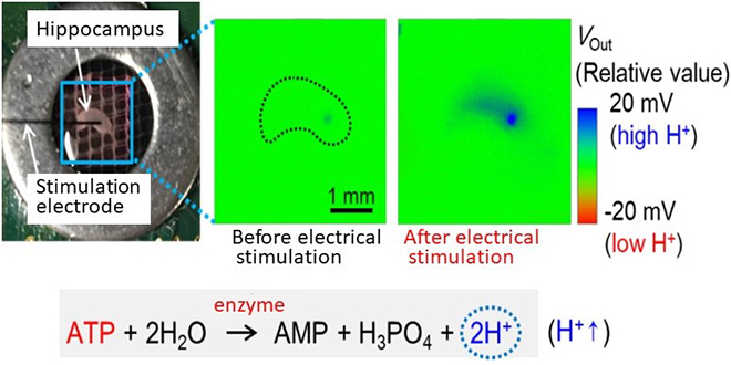
In addition, we found that by chemically treating the surface and by regulating the thickness of the detection membrane to about 100 nm, ATP can be detected at a high sensitivity around the sensing area. After actually attaching a hippocampus to the sensor and conducting biological experiments, the output images changed in accordance with electrical stimulation, and we confirmed that these signals originated from ATP. This is a world-first example of successfully visualizing ATP discharged from brain tissue without the use of fluorescent or other labels.
Since the communication between neurons and glial cells in the hippocampus are presumed to be the elementary process of processing and dispatching information for memory and learning, this research can be expected to contribute to the elucidation of the true mechanisms behind the processing and dispatching of information in the brain. In order to realize this, it is also necessary to achieve imaging and analysis of the temporal and spatial distributions of extracellular neurotransmitters released by neurons and glial cells (which are 10 times greater in number than neurons).
Prospects
The research teams in Toyohashi University of Technology and Yamanashi University succeeded in imaging ATP released from brain tissue. This was achieved using information on ions obtained from a sensor with a spatial resolution of 40 μm, created by improving technology on ATP detection membranes. The research group that developed this sensor at Toyohashi University of Technology, to which I belong, also succeeded in developing an image sensor with a high spatial resolution of 2 μm, approaching the size of a synapse.
By using this sensor with this newly developed ATP detection membrane, it will become possible to visualize ATP released from local areas in cells. In addition, multiple and simultaneous imaging of interactive processes between ATP and other neurotransmitters or different types of ions could bring new information leading to novel insights with physiological significance. Such results could be beyond the scope of those achievable by conventional optical information, such as that from fluorescent microscopes.
This research was supported by Japan Science and Technology Agency (JST) CREST Grant Number JPMJCR14G2.
脳内神経伝達物質を可視化する化学の目を持つセンサ
病気の早期診断や創薬貢献に向けて電気学会 「第35回センサ・マイクロマシンと応用システム」 シンポジウム 五十嵐賞 受賞 (2018年10月)
By 土井 英生
豊橋技術科学大学電気・電子情報工学系の研究チームと山梨大学医学部の研究チームは、生体物質検出膜を修飾した半導体イメージセンサ用いて脳組織切片から放出された神経伝達物質ATP (アデノシン三リン酸 Adenosine Triphosphate) の可視化に成功しました。このセンサは、ATPを認識する酵素を応用することで水素イオンに変換して溶液中のATP拡散を非標識(蛍光標識無し)でリアルタイムに観察可能です。センサ上に、脳組織のような生体組織や臓器を乗せて計測することができ、細胞外応答の2次元分布を画像情報としてダイレクトに得ることが可能です。
本研究の成果を2018年10月に北海道で開催された電気学会の「第35回センサ・マイクロマシンと応用システム」シンポジウムにおいて発表し、五十嵐賞を受賞しました。これは、最も優秀な発表をした35歳以下の若手研究者に贈られるものです。
本研究に取り組んだきっかけ
私がこの研究に取り組んだきっかけは、澤田教授の研究室で開発された”目に見えない化学現象を可視化できる半導体イメージセンサ”に興味を持ったからです。このセンサは私たちの体の中に存在する”イオン”の動きを動画像情報として得ることができます。本センサを応用してこれまで誰も見ることのできなかった脳内の化学現象を可視化するセンサ開発に取組んでいます。また、医学部の先生や研究所の方々と連携して研究を進めるプロジェクトに惹かれたこともきっかけの一つです。現在は、山梨大学医学部 小泉教授の研究グループと共同で医療応用を目指したイメージセンサの開発に取り組んでいます。
研究の苦労話
最も苦労した点は、ATPイメージングに向けて検出膜の成膜技術を検討したことです。約5mm x 5mm のアレイセンサ上でより早く高感度に、そしてきれいにイメージングするために、検出膜材料の物質量や膜厚を追求しました。その結果、疎水性のセンサ表面を薬品処理して滴下溶液の量が少なくても膜厚をコントロールできるようになりました。センサの検出速度に着目して測定データを解析した結果、膜厚を100 nm程度にすることがATP検出に最適であることを実験的に示しました(理論検出限界まで検出感度を向上)。1 sampleの準備から実験結果の解析に30時間を要することも多々ありましたが、山梨大学での生物実験に向けて緻密にデータを取得し、解析することがセンサ性能の向上に繋がりました。
また、山梨大学で生物実験を幾度と重ねる過程では、海馬は培養過程で切片表面にできる細胞の瘢痕(鎧のような層)が原因で神経伝達物質放出の妨げになるのではないかと考え、培養しない急性タイプを提案しました。その結果、海馬自体の明瞭な画像が撮れるようになり、最終的には放出されたATPの画像取得に繋がりました。
受賞に関するエピソード
初めは受賞できてうれしいという率直な気持ちが大きかったのですが、指導教員の澤田教授から、この受賞は本研究グループでは〇〇先生以来20年ぶりの快挙だよ!と伝えられ、名誉のある賞を頂けたんだなと改めて受賞の実感が湧いてきました。また、論文執筆の段階から発表直前まで研究成果発表での話し方や魅せ方を工夫していたので、評価して頂けたことに喜びもひとしおでした。これを糧に今後の研究にも一層注力していきたいと思っています。
研究内容の詳細
脳内において活動的な細胞として注目されているグリア細胞がATPと呼ばれる神経伝達物質を細胞外に放出し、神経細胞の活動(シナプス伝達、シナプスの構造:1-2µm)を制御することで脳機能制御に大きな役割を果たしていることが示されつつあります。そのため、シナプスのような局所部位における細胞外ATPを高い空間分解能でイメージング及び解析することが求められますが、ホタルの光を利用した従来の生物発光法は空間解像度が200µm程度と細胞レベル以上であることからATPの放出部位を可視化できないという問題がありました。
そこで、私たち研究チームは、これまで開発してきた40µmの空間分解能を有するイオンイメージセンサ上にATPを選択的に検出する酵素を修飾したATPイメージセンサを開発しました。本センサは、ATPと酵素の化学反応を用いることにより水素イオンに変換してATPを検出します。また、センサに化学的な表面処理を施し、検出膜を100nm程度に制御するとATPをセンシングエリア近傍で高感度に検出できることがわかりました。そして実際に海馬をセンサ上に密着させて生物実験した結果、電気刺激依存的に出力画像が変化し、ATPに由来した信号であることを確認しました。これは、蛍光標識などのラベルを使用せずに脳組織から滲み出てきたATPの可視化に成功した世界初の例になります。
海馬に集積している神経細胞とグリア細胞によるコミュニケーションは、記憶や学習における情報処理・発信の素過程であることが推測されていることから、本研究は真の情報処理・発信のメカニズム解明に繋がることが期待されます。さらに、神経細胞とその10倍も数の多いグリア細胞による細胞外情報伝達物質の時空間分布をイメージング及び解析することは脳機能の解明に必要不可欠であると考えています。
今後の展望
豊橋技術科学大学と山梨大学の両研究チームは、ATP検出膜の成膜技術を改善することにより、40μmの空間分解能を持つセンサ上でイオン情報を元に脳組織から放出されたATPの画像化に成功しました。センサを開発する私の所属する豊橋技術科学大学の研究グループはシナプスサイズに迫る2µmの高空間分解能を有するイメージセンサも実現しており、このセンサに開発したATP検出膜を応用することで、細胞の局所から放出されるATPを可視化できることが期待されます。さらに、ATPと他の神経伝達物質や種々のイオンなどとの相互作用を複数同時にイメージングすることにより従来の蛍光顕微鏡のような光情報では得ることのできなかった生理学的意義の解明に繋がる新たな情報を得ることが期待されます。
本研究はJST CREST Grant Number JPMJCR14G2の支援を受けて行われました。
Student Profile
| Name | Hideo Doi |
|---|---|
| Affiliation | Department of Electrical and Electronic Information Engineering |
| Title | Doctor course student (1st grade) |
| Fields of Research | Integrated Circuits Devices/Semiconductor Devices |
ここでコンテンツ終わりです。