
ここからコンテンツです。
Intracellular recordings using nanotower electrodes
Nanoscale-tipped high-aspect-ratio vertical microneedle electrodes for intracellular recordings By Takeshi Kawano
Takeshi Kawano and his colleagues have developed an intracellular recording device, which has > 100-µm-long three-dimensional nanoscale-tipped microneedle-electrodes. Moreover, they demonstrated the needles’ penetration into muscle cells and measured the signals. The nanoelectrode, whose size is longer than the conventional intracellular nanoelectrode (< 10-µm long), has the potential to be used in cells that are deep within a tissue, such as cells in brain slices or brain in vivo, thus accelerating the understanding of the brain.
Our current understanding of how the brain works is very poor. The electrical signals travel around the brain and throughout the body, and the electrical properties of the biological tissues are studied using electrophysiology. When measuring the voltage or current across the cell membranes, intracellular recording outperforms extracellular recording in terms of amplitude and quality of neuronal signals. Nanowire-and nanotube-based devices have been developed for the intracellular recording applications to demonstrate the advantages of these devices having high spatial resolution and high sensitivity.
However, the length of these nanowire/nanotube electrode devices is currently limited to less than 10 µm due to process issues that occur during fabrication of high-aspect-ratio nanoscale devices, which are more than 10-µm long. Thus, conventional nanodevices are not applicable to neurons/cells within thick biological tissues, including brain slices and brain in vivo.
A research team in the Department of Electrical and Electronic Information Engineering and the Electronics-Inspired Interdisciplinary Research Institute (EIIRIS) at Toyohashi University of Technology has developed three-dimensional microneedle-based nanoscale-tipped electrodes (NTEs) that are longer than 100 µm. The needle length exceeds that of the conventional nanowire/nanotube-based intracellular devices, thus expanding the range of applications of nanodevices in intracellular recording, such as deep tissue penetration. Additionally, they perform intracellular recordings using muscle cells.
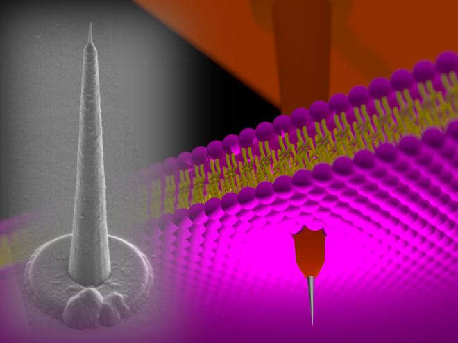
“A technological challenge in electrophysiology is intracellular recordings within thick biological tissue. For example, a needle length of more than 40 µm is necessary for performing brain slice experiments. However nanoscale diameter needles with a high-aspect-ratio have a long hair-like nanostructure which makes it almost impossible to penetrate thick tissue due to insufficient stiffness. On the other hand, our NTE, which is a 120-µm-long cone-shaped electrode, has sufficient stiffness to punch tissues and cells”, explains the first author PhD candidate, Yoshihiro Kubota.
The leader of the research team, Associate Professor Takeshi Kawano said "Although we demonstrated the preliminary results of our NTE device, the batch fabrication of such intracellular electrodes, which have a needle length more than 100 µm, should lead to an advancement in device technologies. This will eventually lead to the realization of multisite, depth-intracellular recordings for biological tissues, including brain slices and brain in vivo, which are beyond the capability of conventional intracellular devices.”
As addressed by the research team, the NTE has the potential to be used in cells that are deep within a biological tissue, including brain slice and brain in vivo, thus accelerating the understanding of the brain.
This study was supported by
- Grants-in-Aid for Scientific Research (S) (No. 20226010) and (A) (No. 25249047) from JST
- Grants-in-Aid for Young Scientist (A) (No. 26709024) and (B) (No. 22760251) from JST
- The PRESTO Program from JST
- Leading Graduate School Program R03 of MEXT
- Grant-in-Aid for Scientific Research (C) (No. 24590350) from JST
- the Asahi Glass Foundation and the Takeda Science Foundation
Reference
Yoshihiro Kubota, Hideo Oi, Hirohito Sawahata, Akihiro Goryu, Yoriko Ando, Rika Numano, Makoto Ishida, and Takeshi Kawano (2016). Nanoscale-tipped high-aspect-ratio vertical microneedle electrodes for intracellular recordings, Small, Article first published online: 8 April 2016 | DOI: 10.1002/smll.201600172
細胞内電位計測のための‘ナノタワー’電極デバイス
細胞内電位計測のための高アスペクト比・ナノスケール先鋭化マイクロニードル電極河野剛准教授らは、細胞内電位計測応用として、長さが100 µm以上、先端の直径がナノサイズに先鋭化された3次元的マイクロプローブ電極デバイスを開発した。また、彼らは開発した電極を用いたマウスの筋細胞への電極刺入及び細胞内からの信号計測を実証した。研究グループが開発したナノ電極は、長さが10 µm程度以下に留まっていた既存のナノ電極デバイスの長さを大きく上回るもので、脳スライスや生体内(in vivo)計測での組織深部の細胞における細胞内電位計測の可能性を拡大し、脳計測技術さらには私たちの脳の理解を加速させるツールとして今後期待される。
脳に対する私たちの理解は未だもって不十分であり、今後の脳計測のさらなる進展が必要である。その中でも電気生理学的手法は脳組織の神経回路網を理解するための有力な方法である。細胞外電位計測と比較すると、細胞内電位計測は信号の電圧値の大きさや信号の質(シナプス後電位等)などの点において優れた方法である。既存のガラス管電極の空間分解能や感度を上回る技術として、ナノテクノロジーを駆使したナノワイヤやナノチューブによる細胞内電位計測用のデバイスが近年開発されてきている。
しかしながら、これらのナノ電極デバイスは製作手法の制約からその電極長が10 µm以下に留まっている。そのため、これら従来のナノデバイスでは、脳スライスや生体内(in vivo)脳組織などの厚さのある生体組織の深部に位置する細胞への応用が困難であった。
豊橋技術科学大学の電気・電子情報工学専攻の学生・研究者たちとエレクトロニクス先端融合研究所の研究者たちは長さ100 µmを超える三次元的ナノスケール先鋭化マイクロニードル電極(Nanoscale-tipped electrode, NTE)デバイスを開発した。開発した電極ニードルの長さは、従来の細胞内電位計測用ナノ電極の長さを大きく上回り、ナノデバイスによる細胞内電位応用の応用範囲を大きく広げる。彼らはまた、開発したNTEによる筋細胞への電極の刺入及び信号計測を実証した。
「従来のナノ電極を用いた電気生理学的手法における技術的な課題は、組織深部の細胞に対する細胞内電位計測である。例えば脳スライスの場合、損傷の少ない細胞は、切片表面から深さ約40 µm以上に位置する。しかし、40 µm長の高アスペクト比のナノ電極では、そのナノ構造の不十分な剛性のために刺入が困難であった。一方で、私たちが開発した120 µm長のNTEは、その円錐に近い電極形状により、細胞や組織を貫くのに十分な剛性を実現できる」と筆頭著者である博士後期課程の久保田吉博は説明する。
本研究のチームリーダーである河野剛士准教授は「今回は、提案するNTEデバイスの基礎的な特性結果を示したに過ぎないが、長さ100 µm以上のナノ電極を一括製作が可能な今回の技術は、細胞内用のデバイス技術をさらに発展させるもので、また本提案デバイスにより最終的にはこれまでの細胞内計測では困難とされてきた脳スライスやin vivo(生体内)脳組織を含む組織深部での多点、同時における細胞内電位計測の実現が可能性である」と考えている。
今回開発されたNTE電極は、脳スライスや生体内(in vivo)脳組織などの組織深部における細胞内電位計測の可能性を拡大させるもので、脳計測技術さらには私たちの脳の理解を加速させるツールとして今後期待される。
本研究は、以下の助成を受けて行われました。
- 文部科学省・日本学術振興会科学研究費基盤研究S(20226010)、基盤研究A(25249047)、若手研究A(26709024)、若手研究B(22760251)
- 科学技術振興機構さきがけ(PRESTO)
- 文部科学省・日本学術振興会博士課程教育リーディングプログラム(R03)
- 文部科学省・日本学術振興会科学研究費基盤研究C(24590350)
- 公益財団法人武田科学振興財団、公益財団法人旭硝子財団
Researcher Profile
| Name | Takeshi Kawano |
|---|---|
| Affiliation | Department of Electrical and Electronic Information Engineering |
| Title | Associate Professor |
| Fields of Research | Micro/Nano Devices, Neural Interface Devices |
ここでコンテンツ終わりです。