
Kobayashi, Masakazu

| Affiliation | Department of Mechanical Engineering |
|---|---|
| Title | Professor |
| Fields of Research | Analysis and evaluation of material microstructure / X-ray imaging |
| Degree | Dr. Eng. (Utsunomiya Univ.) |
| Academic Societies | The Japan Institute of Metals / The Japan Institute of Light Metals / Japan Fundary Engneering Society / The Iron and Steel Institute |
| m-kobayashi@me Please append ".tut.ac.jp" to the end of the address above. |
|
| Laboratory website URL | http://ma.me.tut.ac.jp |
| Researcher information URL(researchmap) | Researcher information |
Research
My research is development of the 3-D imaging technology by means of synchrotron and laboratory scale X-ray CT devices, and its application to the materials science and engineering. It is expected that high-resolution 3-D visualization of microstructure in metal materials make deformation, damage, fracture and fatigue mechanisms clear. Furthermore, my laboratory develops the technique that measures high-densely dynamics information (stress, strain, displacement, crack driving force and so on) in 3-D by a tracking of microstructural features inside materials. So we try to link quantity data extracted from 3-D image to various theories, a model, an experience-based rule, a numerical analysis that have been built till now.
Theme1:Characterization of microstructure in materials by high-resolution X-ray CT
Overview
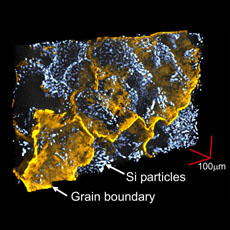 Fig. Grain-boundary and Si particles distribution in AC4CH aluminum cast alloy
Recently, characterization of complex microstructure in various materials is available by X-ray computed tomography (CT) because spatial resolution is improving in the X-ray CT that utilizes synchrotron radiation generated from high flux source. In this study, effective quantitative evaluation methods that measure various microstructures of materials in X-ray CT are developed. For example, we can measure volume fraction, distribution and shape in defects (like pores) and particles (like precipitates and dispersion). Furthermore, the technique to visualize grain-boundary position in 3D is also developed by penetrating liquid metals into grain-boundary and improving detectability with utilizing X-ray refraction contrast.
[Publications]
◆M. Kobayashi, Y. Dorce, H. Toda, H. Horikawa, Effect of local volume fraction of microporosity on tensile properties in Al-Si-Mg cast alloy, Materials Science and Technology, Vol. 26, No. 8, 962-967, 2010
◆M. Kobayashi, H. Toda, K. Uesugi, T. Ohgaki, T. Kobayashi, Y. Takayama, B.-G. Ahn, Preferential penetration path of gallium into grain boundary in practical aluminium alloy, Philosophical Magazine, 86(28),4351-4366, 2006
Keywords
Theme2:Development of three-dimensional high-accurate strain mapping by using high-resolution X-ray CT
Overview
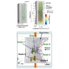 Fig. 3D strain mapping (upper), 3D image of grains by liquid metal wetting (under)
In case of X-ray CT observation in metals for structural materials in the third generation synchrotron radiation facility, SPring-8, several hundreds of thousands of micro-pores and particles are seen in a material. We can obtain and visualize 3D strains in the inside of materials by analyzing displacement of markers that are tracked during deformation, because these microstructural features (we can use micro-pores and particles as markers) move with obeying local deformation during tensile test or compression test (upper figure (a) and (b));. However, no error tracking is difficult in several hundreds of thousands of markers in three-dimensional space. In this study, tracking algorithms are developed to obtain high-dense and high-accurate strain mapping. If such tracking is available, we can investigate and understand local deformation mechanism in the inside of materials. Moreover, with combining strain mapping with the grain-boundary visualization technique applying liquid metal wetting into grain-boundary, grains-deformation analysis in polycrystalline metals is also available (under figure). Conventional microscope can access the information on only surface. If we utilize X-ray CT, which is non-destructive inspection, we can understand a factor that affects mechanical properties directly because we can see deformation and fracture in the inside of materials. If the true deformation and fracture mechanism, which we can't understand by surface observation, are found, we can design the materials which are hard to be broken more.
[Publications]
◆M. Kobayashi, H. Toda, Y. Kawai, T. Ohgaki, K. Uesugi, D.S. Wilkinson, T. Kobayashi, Y. Aoki, M. Nakazawa, “High-density three-dimensional mapping of internal strain by tracking microstructural features”, Acta Materialia, 56(2008), 2167-2181
◆M. Kobayashi, H. Toda, Y. Kawai, T. Kobayashi, K. Uesugi, David S. Wilkinson, Eric Maire, Y. Aoki, 'Measurement of 3-D strain distribution by means of high- resolution X-ray CT image and tracking of microstructural features ', Journal of Japan Institute metals, 71(2),181-186, 2007
Keywords
Title of class
Introduction to Materials Engineering / Differential Equation / Reliability Engineering for Materials/ Experimental Practice for Mechanical Engineering / Intro. Materials Science/