
ここからコンテンツです。
Automated detection of isolated single cells using microscope images and AI
Contributing to the development of new treatments with single-cell analysis and isolation Moeto Nagai and Tanmay Debnath
A research team, led by Professor Moeto Nagai and comprised of researchers from the Department of Mechanical Engineering and Institute for Research on Next-generation Semiconductor and Sensing Science (formerly known as the Electronics Inspired Interdisciplinary Research Institute (EIIRIS),Toyohashi University of Technology, has successfully used AI to achieve single-cell isolation. The method involves using microwells to isolate single cells and then applying deep learning to the microscopic images containing single cells in the microwells. The machine learning model prepared by the team makes it possible to automatically detect single cells in microscopic images and reduce human effort.The acquisition of a large volume of single-cell data allows researchers to efficiently investigate the characteristics and functions of individual cells, which can lead to the establishment of new treatment methods.
A cell is the most basic unit of life, and elucidation of cell characteristics can contribute to a better understanding of diseased cells and thus to the development of new treatment methods. There has been a growing interest in developing methods of isolating single cells to study their functions. However, the physical isolation of single cells requires cell patterning tools. In addition, as the detection of single cells often relies on human eyes and classification, the required human effort has created a bottleneck that has hindered the acquisition of data.
Initially, the research team developed a method of isolating and trapping single cells in microwells of 30μm diameter formed in a hydrogel patterned by optical patterning. The micro-patterned hydrogel offers the advantages of convenience and stability. These microwell structures allowed the research team to successfully isolate single human cells from a cell suspension. As hydrogel is highly biocompatible and can withstand a long incubation time in a cell medium, it was possible to extend the observation period of cell behavior.
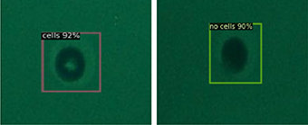
Presence of single cell (left) and absence of single cell (right)
Next, the research team classified the cells trapped in microwells of 30μm diameter in terms of the presence or absence of single cells and used the images as training data to perform deep learning. The object detection model resulting from the learning could then be used to detect the presence of single cells based on input images. This made it possible to predict the presence of single cells with a mean Average Precision (mAP) of 0.989 (the higher the better) and an average inference time of 0.06 seconds (the shorter the better). This algorithm offers high detection accuracy while reducing experiment time, which leads to improving high-throughput single-cell analysis.
At first, the research team used bright-field microscopy images as input datasets, which are commonly used for the observation of single cells, but as these images did not offer high contrast, there were limits to the improvement of the detection capability. Performance stagnated at an mAP of 0.801 and an average inference time of 0.09 seconds. The team then switched to cells stained with fluorescent dyes and the use of fluorescence microscopy images as the input datasets, which allowed them to achieve convergence at an mAP of 0.989 after 1,200 epochs of training. This indicates that preparing high-contrast input datasets, which make it easier for humans to detect the presence of the cells, is also important for the use of AI.
Research team leader Moeto Nagai explains: "We wanted to apply AI to the detection of single cells. As I had been performing mostly experiment-based research, the use of experimental data for AI research seemed to me to be a significant obstacle. However, the participation of graduate student Tanmay Debnath, the lead author of our study, who has experience in the research and development of AI technology, meant that we could rapidly make use of AI and ultimately led to the success of our development."
The single-cell isolation and detection developed by this research can also be used to automatically monitor the activities of single cells. This research achieves accurate and highly reliable automated cell detection, while reducing human labor. Future applications for single-cell analysis include medical engineering applications in a wide range of areas such as cancer diagnosis, immune response, and drug discovery screening, which will contribute to the discovery of new treatment methods.
Reference
Tanmay Debnath, Ren Hattori, Shunya Okamoto, Takayuki Shibata, Tuhin Subhra Santra, Moeto Nagai (2022). Automated detection of patterned single-cells within hydrogel using deep learning, Scientific Reports volume 12, Article number: 18343, DOI: https://doi.org/10.1038/s41598-022-22774-0
AIで顕微鏡画像から分離した単一細胞を自動検出する
単一細胞解析や分離による新規治療法の確立に向けて永井 萌土、タンメイ デブナス
豊橋技術科学大学機械工学系と次世代半導体・センサ科学研究所(旧称:エレクトロニクス先端融合研究所)の研究チーム(永井萌土教授ら)は、マイクロウェルで単一細胞を分離後、ウェル内の単一細胞を含む顕微鏡画像に深層学習を適用して、AIを用いた単一細胞検出を実現しました。構築した機械学習モデルにより人の関与を減らしながら、顕微鏡画像から自動的に単一細胞を検出できます。単一細胞データの大量取得により、細胞個々の特徴や機能を効率的に調査することができ、新規治療法の確立につながります。
細胞は生命の最も基本的な単位で、細胞の特性解明は、細胞の病気の理解や新規治療法の確立につながります。単一細胞を分離し、機能を調査するための手法の開発に強い関心が寄せられています。ただし単一細胞を物理的に分離するには、規格化する道具が必要です。さらに単一細胞の検出は、人間の目と判断で行われることが多く、人間の関与が律速となり、データの取得を妨げていました。
最初に、光パターニングで作製した直径30µmのハイドロゲルを用い、内部に単一細胞を分離して捕獲する方法を開発しました。微細加工のフォトリソグラフィーで規格化するハイドロゲルは、簡便かつ安定な利点を持ちます。このマイクロウェル状の構造体により、細胞懸濁液からヒト単一細胞を分離捕獲しました。ハイドロゲルは生体適合性も高く、細胞培地中での長い培養時間に耐えることができ、細胞の挙動を観察する時間も延長できました。
続いて、サイズ30µmのマイクロウェルアレイ内の単一細胞を分類し、細胞ありとなしの画像を教師データとして深層学習を行いました。学習済みの物体検出モデルは、入力画像を元に単一細胞を検出するようになりました。平均適合率0.989(1.0に近いほど良い)、平均推論時間0.06秒(短いほど良い)で単一細胞の存在を予測するようになりました。このアルゴリズムは、高い検出精度かつ実験時間の短縮を可能にし、ハイスループットな単一細胞分析の強化につながるものです。
最初は、入力データに一般的に用いられる観察法である明視野顕微鏡の画像を用いていましたが、コントラストが高くないこともあり、検出能力の向上に限界がありました。性能は平均適合率0.801、平均推論時間0.09秒に留まりました。ここで細胞を蛍光染色し、入力データを蛍光顕微鏡画像に変更したところ、上述の平均適合率0.989に1200エポックで収束させることができました。このことは、入力データにコントラストが高く、人間でも見つけやすいデータを用意することが、AIにとっても重要なことを示しています。
研究チームのリーダーである永井萌土は「単一細胞の検出に対し、AIのアプローチを適用したいと考えていました。私は実験系の研究を中心に進めていたことから、実験データをAIの研究に展開することにハードルの高さを感じていました。ここでAI技術の研究開発を行ってきた、筆頭著者で大学院生のTanmay Debnathさんがプロジェクトに参画したことにより、AIの利用を迅速に進められ、開発の成功につながりました」と説明しています。
本研究で開発した単一細胞の分離と検出は、単一細胞の活動の自動観察に用いることができます。本研究により、人間の労力を減らしながら、正確で信頼性の高い細胞の自動検出をすることができます。今後は、シングルセル解析として、がん診断、免疫応答、創薬スクリーニングなどの種々の医用工学応用に展開し、新しい治療法の発見にも結びつけていきます。
Researcher Profile

| Name | Moeto Nagai |
|---|---|
| Affiliation | Institute for Research on Next-generation Semiconductor and Sensing Science (formerly known as Electronics Inspired Interdisciplinary Research Institute) |
| Title | Professor |
| Fields of Research | BioMEMS / Biohybrid System / Micro-Nano Mechatronics / MicroTAS / Biofabrication / Micromachining |
ここでコンテンツ終わりです。