
Visualization of chemical phenomena in the microscopic world using semiconductor image sensors
Aiming at the elucidation of brain functions and medical applications Hideo Doi
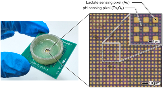
A Toyohashi University of Technology research team led by Professor Kazuaki Sawada and Project Assistant Professor Hideo Doi of the Department of Electrical and Electronic Information Engineering, has developed a semiconductor sensor that enables real-time observation of the dynamics of two types of biomolecule in solutions. By using semiconductor technology to pattern a thin metal film functioning as a neurotransmitter-sensitive membrane on sensor pixels arranged two-dimensionally in a 2 µm pitch, the sensor captures the movement of hydrogen ions and lactate (neurotransmitters) in a solution as image data. The accuracy level achieved wasto the degree of a temporal resolution of milliseconds and a spatial resolution of several microns (approximately 1/17 the thickness of a strand of hair) were achieved. The system is expected to measure the relationship between the distribution of multiple types of neurotransmitters and ions that vary in time and space between cells with high spatiotemporal resolution.
The brain is essentially made up of around 100 billion neurons, accompanied by ten times as manynon-neuronal cells (Glial cells). Action potentials generated by these neurons are transmitted between cells as electrical signals. On the other hand, it is known that chemical substances or ions called neurotransmitters move between the tiny voids at the cell-cell junctions and play an important role both in controlling brain functions as chemical signals, as well as in the pathophysiology of the brain. In other words, in order to investigate detailed brain functions and pathologies based on the elucidation of chemical signal transduction mechanisms, it is necessary to develop technology to visualise and measure the dynamics of chemical substances distributed in the extracellular space from a few micrometres (cellular level) to several hundred µm (cell population level). However, most conventional devices for chemical signal detection tend to rely on passing an electric current through a single tiny electrode with a diameter of several tens of microns, or electrodes arranged at intervals of several 100 microns.These barriers to miniaturization and downsizinghave, up to now, prevented the realization of sensors capable of measuring the distribution of chemical information at the micron level.
Professor Sawada's laboratory however, has developed a semiconductor image sensor that captures ion movement like a camera. It achieves this by using semiconductor technology to miniaturise and integrate potential detection elements that are sensitive to hydrogen ions, which has promoted the development of highly functional sensors. The researchers fabricated an imaging device for multisensing in which metal electrodes are formed in a lattice shape at intervals of 6 µm on the developed sensor array, and realized the real-time simultaneous measurement of lactate and hydrogen ions involved in memory formation by applying a recognition element (enzyme) that selectively detects biomolecules. In addition, recognition elements corresponding to biomolecules can be also patterned on the electrode array, enabling the simultaneous multichemical measurement of two or more types of neurotransmitters.
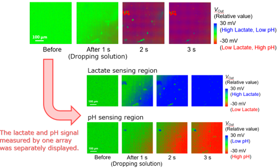
In the past, one type of biomolecule was measured with a single-point electrode, or one to three types of biomolecules were measured with an electrode array sensor with low spatial resolution. The newly developed feature of “capturing, visualizing, and measuring” two types of chemicals on a surface with high spatial resolution enables the detection of chemical signals in microscopic regions, such as between cells, along with spatiotemporal analysis. This advancement offers insights into chemical phenomena that were previously inaccessible using point-based sensors. In a collaborative experiment with researchers from the Faculty of Medicine, University of Yamanashi, experts in brain science and pharmacology, the response of hippocampal cells (responsible for memory) to drug stimulation was observed. The researchers simultaneously measured lactate release and extracellular pH. This breakthrough enables direct placement and measurement of cells and tissues without any labeling, allowing observation of spatiotemporal changes in ions and neurotransmitters in biological samples. Building on this world-first ion image sensor technology, the team aims to advance its development for social implementation and medical applications.
We plan to proceed with the further enhancement of the functionality and performance of sensors to expand the range of substances to be measured. Furthermore, using sections of diseased tissue, such as brain tissue and tissue forming solid tumours, we plan to undertake applied measurements to elucidate the physiological significance of how molecular dynamics in the environment outside the cell changes in time and space. We also plan to apply this technology to analyzing interactions among chemical substances in the complex extracellular microenvironment and to verify their pharmacological effects.
Reference
Hideo Doi, Hayato Muraguchi, Tomoko Horio, Young-Joon Choi, Kazuhiro Takahashi, Toshihiko Noda, Kazuaki Sawada, “Real-time simultaneous visualization of lactate and proton dynamics using a 6-µm-pitch CMOS multichemical image sensor”, Biosensors and Bioelectronics, Vol.268, 116898. https://doi.org/10.1016/j.bios.2024.116898
半導体イメージセンサでミクロな世界の化学現象を可視化する
脳機能の解明や医療応用を目指して 土井 英生豊橋技術科学大学 電気・電子情報工学系の澤田和明教授、土井英生特任助教らの研究チームは、液中で異なる2種類の生体分子の動態をリアルタイムに観察できる半導体センサを開発しました。このセンサは2 µm間隔で2次元に配置されたセンサ画素上に神経伝達物質感応膜として機能する金属薄膜のパターンが半導体技術で格子状に形成されており、水素イオンと乳酸(神経伝達物質)が液中で実際に動いていく様子を画像情報として取得することが可能です。ミリ秒の時間分解能と数ミクロンの空間分解能(髪の毛の約1/17サイズ)を達成し、細胞間で時間・空間的に変化する複数種類の神経伝達物質やイオンの分布の関係を高い時空間分解能で計測することが期待できます。
脳内には1000億個の神経細胞とその10倍の非神経細胞(グリア細胞)が存在しており、細胞が発する活動電位が電気信号となり情報が伝達されています。一方、細胞と細胞のつなぎ目には微小な空隙が存在し、その間を神経伝達物質と呼ばれる化学物質またはイオンが移動することで化学信号として脳機能制御に重要な役割を果たすことや脳の病態生理に深く関わることなどが知られています。つまり、化学シグナルの伝達機構解明に基づく脳の詳細な機能や病態を調べるためには、数ミクロン(細胞レベル)~数100 µm(細胞集団レベル)の細胞外空間に分布する化学物質の動態を可視化計測する技術開発が求められます。しかし、従来の化学信号計測デバイスのほとんどは、直径数十ミクロンの微細な単一電極や数100ミクロン間隔に電極を並べて、電流を流すタイプのために微細化・小型化が難しくミクロンレベルで化学情報の分布を計測するセンサは実現されていませんでした。
一方、澤田教授の研究室では、水素イオンに感応する電位検出素子を半導体技術で微細化・集積化することでイオンの動きをカメラで撮影するように見ることのできる半導体イメージセンサを開発し、センサの高機能化を進めてきました。開発した半導体イメージセンサ上に金属電極を6 µm間隔で格子状に形成し、生体分子を選択的に検出する認識素子(酵素)を応用することで記憶形成に関わる乳酸(Lactate)と水素イオンのリアルタイム同時計測を実現しました。また、生体分子に対応する認識素子を電極上に塗分けることもできるため、2種類以上の神経伝達物質のマルチ計測も可能になります。
従来は1種類の生体分子を1素子で計測するか、1~3種類の生体分子を空間分解能の低い電極アレイセンサで計測されていました。一方で、本研究チームが開発した高い空間分解能で2種類の化学物質を“面で捉え可視化計測できる”特徴は、細胞間のようなミクロな空間領域で変化する化学信号の時空間解析を可能とし、これまでの“点”で計測するセンサでは得られなかった化学現象のメカニズム解明に貢献することが期待できます。共同研究を進めている脳や薬理学に詳しい山梨大学医学部の研究者らと行った脳組織(海馬:記憶を司る)の計測実験では、薬剤刺激に細胞が応答し、乳酸が放出される様子と細胞外pHを同時に可視化することに成功しました。本センサは、細胞や組織を直接乗せて測ることができ、生体試料における神経伝達物質の時空間変化を、顕微鏡を覗くようにして観察することができるようになると考えており、世界に先駆けて開発・応用してきたイオンイメージセンサ技術をさらに発展させ、社会実装・医療応用を目指して取り組んでまいります。
計測対象物質の拡大に向けたセンサのさらなる高機能化、高性能化を進める予定です。さらに、脳の組織や固形腫瘍を形成するような病変組織の切片を用いて、細胞の外の環境における分子動態が時間・空間的にどのように変化するか、その生理学的な意義の解明に向けた応用計測に取り組む予定です。また、細胞外微小環境において複雑に絡み合う化学物質の相互作用解析や薬理効果の検証にも役立てていきたいと考えています。
Researcher Profile
| Name | Hideo Doi |
|---|---|
| Affiliation | Department of Electrical and Electronic Information Engineering |
| Title | Project assistant professor |
| Fields of Research | Electronic device / Biosensor |