
Research highlights
Bio-imaging offers insights into the relationship between circadian and ultradian rhythms.
Living things have rhythms—for example, body temperature rhythms and segmentation clock. In the late 1990s, several clock genes were cloned to elucidate the functions and interactions of rhythms. The feedback loop of transcriptional factor with 24 h period in the suprachiasmatic nuclei (SCN) was proposed to work as a circadian central oscillator, as well as in peripheral tissues including cartilage and bone. On the other hand, the fundamental architecture of skeletal patterning is regulated by ultradian clocks that undergo cycles more than once every 24 hours in embryonic development. In 1997, the oscillatory expression of c-hairy1—a Notch effecter gene—was identified in chick embryos and matched the period of somite formation (every 90 minutes in chicks), called the segmentation clock.
Somitogenesis is one of the most evident events in an ultradian manner, which endows basic repetitive patterns of axial skeleton and its associated tissues during embryonic development. Long bone growth and bone metabolism also exhibit periodic activities in a circadian fashion. Core loops of circadian clock genes are also at work in bone and cartilage.
Here, collaborators at Toyohashi University of Technology and Tokyo Medical and Dental University propose bio-imaging methodology to observe both clocks. Bio-imaging detecting of luminescent and fluorescent signals enables observation of more comprehensive sets of genes and spatio-temporal regulation of these clockwork machineries during development.
In this review paper, the authors also describe the potential of three dimensional imaging for bone research. Topics covered include molecular clocks in skeletal biology and medicine, and how fluorescence imaging would contribute to widening our knowledge of biomedical science.
- Tadahiro Iimura*1, Ayako Nakane1, Mayu Sugiyama1, Hiroki Sato1, Yuji Makino, Takashi Watanabe1 , Yuzo Takagi1, Rika Numano*2 , Akira Yamaguchi*1
- *to whom correspondence should be addressed
- A fluorescence spotlight on the clockwork development and metabolism of bone
- Journal of Bone and Mineral Metabolism On line July 2011
- DOI: 10.1007/s00774-011-0295-3
- 1International Research Center for Molecular Science in Tooth and Bone Diseases, Tokyo Medical and Dental University, Sections of Oral Pathology
- Website: http://www.tmd.ac.jp/dent/opat/opat-J.htm
- 2The Electronics-Inspired Interdisciplinary Research Institute (EIIRIS), Toyohashi University of Technology
- Website: http://www.eiiris.tut.ac.jp/
Rika Numano

Enlarge Image
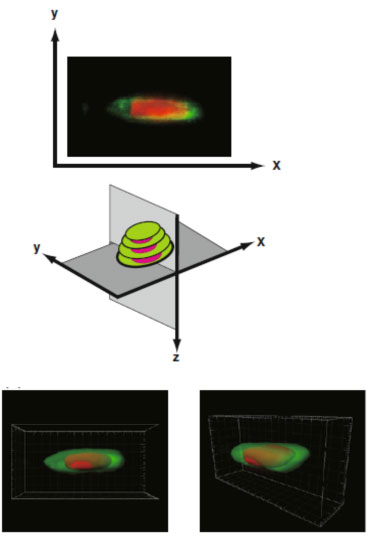
Figure caption: Quantitative 3-dimensional fluorescent imaging on bone tissue
