
Research highlights
Brain regions sensitive to facial color processing
Facial color provides important clues to recognize a person’s emotion and health, and is therefore facial color contains important information for social communication.
Previous research published by Tetsuto Minami and colleagues at the Electronics-Inspired Interdisciplinary Research Institute (EIIRIS) at Toyohashi Tech based on electroencephalography (EEG) has shown that the face-sensitive event related potential (ERP) component (N170) is modulated by facial color, which suggests that face color is important for face detection (Minami et al. Neuroscience, 176, 265-73, (2011)). Moreover, the sensitivity of N170 was found at the left occipito-temporal site (Nakajima et al., Neuropsychologia, 50, 2499-505, (2012)).
However, it is not clear which region of the brain is involved in facial color processing because spatial resolution of EEG is not sufficient.
Here, Tetsuto Minami and colleagues at Electronics-Inspired Interdisciplinary Research Institute (EIIRIS) at Toyohashi Tech, report on the brain regions sensitive to color information for face processing.
The present study aimed to identify the brain regions related to facial color processing by using functional magnetic resonance imaging (fMRI). The researchers measured the brain activity from 25 participants during the presentation of natural- and bluish-colored faces and other scrambled images. Face-selective regions of interest (ROIs) were identified separately for each subject and hemisphere of the brain.
As a result, the bilateral fusiform face (FFA) area and occipital face area (OFA) were identified as brain areas that were activated more to natural-colored faces than to natural-colored scrambled images. The ROI analysis showed that the left FFA was sensitive to facial color, whereas the right FFA was not. Furthermore, the right and left OFA were insensitive to facial color.
In combination with previous EEG studies by this group, the present study showed that the left FFA is related to facial color processing.
- Reference:
- Authors: Kae Nakajima1, Tetsuto Minami2,*, Hiroki C. Tanabe3,4, Norihiro Sadato3,5 and Shigeki Nakauchi1
- Title of original paper: Facial color processing in the face-selective regions: An fMRI study
- Journal, volume, pages and year: Human Brain Mapping Early View (Online Version of Record published before inclusion in an issue)
- Digital Object Identifier (DOI): 10.1002/hbm.22535
- Affiliation(s): 1Department of Computer Science and Engineering, Toyohashi University of Technology, Toyohashi, Aichi, Japan, 2Electronics-Inspired Interdisciplinary Research Institute, Toyohashi University of Technology, Toyohashi, Aichi, Japan, 3Department of Cerebral Research, National Institute for Physiological Sciences, Okazaki, Aichi, Japan, 4Department of Social and Human Environment, Graduate School of Environmental Studies, Nagoya University, Nagoya, Japan, 5Department of Physiological Sciences, the Graduate University for Advanced Studies, Okazaki, Aichi, Japan
- Website: http://www.eiiris.tut.ac.jp/

Tetsuto Minami
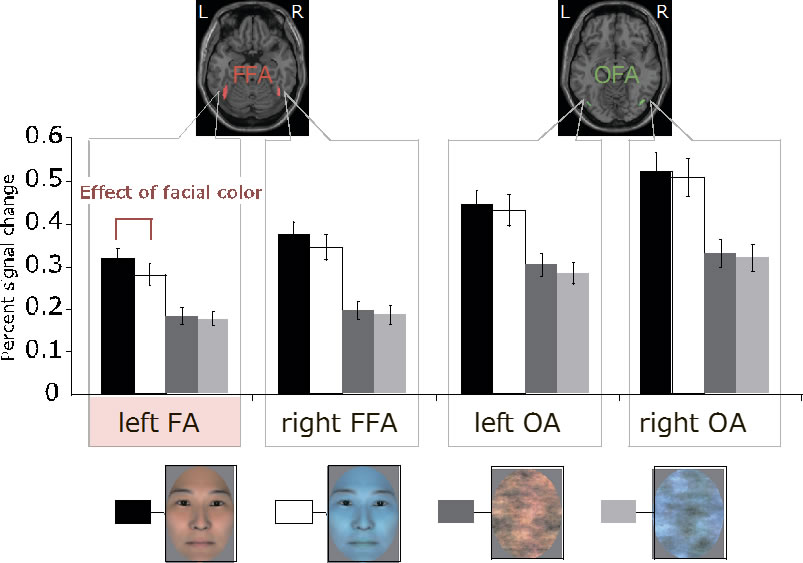
Fig.1: Results of the ROI analyses, which suggests that the left FFA was sensitive to facial color.
Enlarge Image