
Research highlights
Hollow bioprobe: Innovative analytical tool for the life sciences and biotechnology
A deep understanding of the functions of cells is crucial for such as medical diagnostics, drug discovery, and tissue engineering.
With the aim of opening up new possibilities for revolutionary approaches to analyze cellular functions at the single-cell level, Takayuki Shibata and colleagues at Toyohashi University of Technology propose the ‘bioprobe’ : an innovative probe for atomic force microscope (AFM) that is integrated with a sharp-tipped hollow silicon dioxide (SiO2) nanoneedle instead of a conventional solid tip.
The bioprobe enables both the quantitative introduction of the desired biomolecules (DNA, proteins, etc.) into living cells and extraction of an extremely small number of biomolecules or cellular components expressed inside the cells as well as a variety of functions of conventional AFM functions.
The electrically insulating and optically transparent properties of the hollow structure of the SiO2 nanoneedle provides other useful functions including (1) scanning ion conductance microscopy (SICM) for non-damaging imaging of biological cells; (2) scanning near-field optical microscopy (SNOM) for high-resolution optical imaging; and (3) surface enhanced Raman scattering (SERS) spectroscopy for characterizing molecular interactions in the biological cell.
Importantly, bioprobe has the potential to enable simultaneous imaging of multiple signals from single living cells. The researchers are confident that this approach will provide a method for correlation analysis of cellular functions with high spatial and temporal resolution for the development of a viable strategy for improving health and quality of life.
- Reference:
- Authors: Takayuki Shibata, Kenji Nakamura, Shuhei Horiike, Moeto Nagai, Takahiro Kawashima, Takashi Mineta, and Eiji Makino.
- Title of original paper: Fabrication and characterization of bioprobe integrated with a hollow nanoneedle for novel AFM applications in cellular function analysis.
- Journal, volume, pages and year: Microelectronic Engineering 111, 325-331 (2013).
- Digital Object Identifier (DOI): 10.1016/j.mee.2013.02.051
- Affiliations: Department of Mechanical Engineering, Toyohashi University of Technology.
- Department website: MEMS/NEMS Processing Laboratory
Professor Takayuki Shibata
Enlarge Image
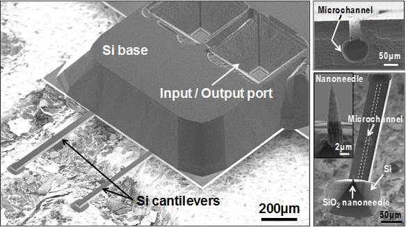
Figure 1: Bioprobe with a fully integrated sharp-tipped hollow SiO2 nanoneedle and fluidic microchannel embedded into a Si cantilever beam structure.
Enlarge Image
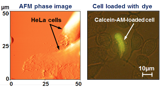
Figure 2: Atomic force microscope (AFM) phase images of HeLa cells in a culture medium and introduction of a fluorescent dye into a living HeLa cell by penetration of its cell membrane with the nanoneedle tip of bioprobe.
Enlarge Image