
ここからコンテンツです。
Super small needle technology for brain
Dissolvable material expands opportunities of flexible microneedles for brain penetrations By Takeshi Kawano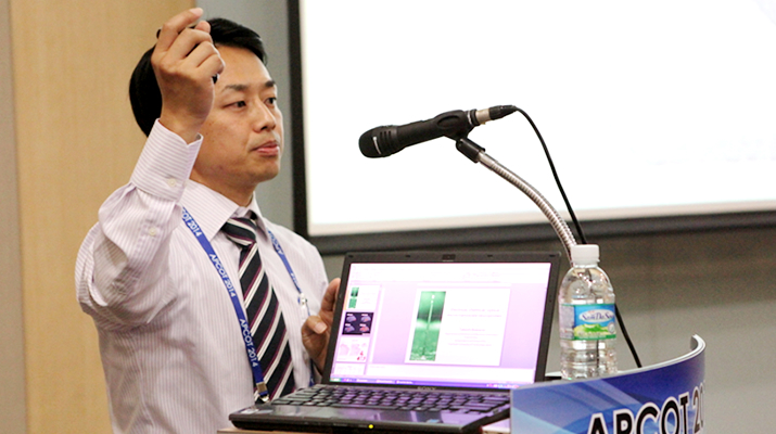
The interdisciplinary collaborative research team led by Associate Professor Takeshi Kawano has now developed a methodology for brain penetration of sub-5μm diameter flexible needles. This should further reduce invasiveness and provide safer tissue penetrations than conventional approaches.
Microscale needle-electrode array technology has been enhancing brain sciences and engineering such as electrophysiological studies, drug and chemical delivery systems, and optogenetic applications.
However, one enduring challenge is to reduce the tissue/neuron damage associated with needle penetration, particularly for chronic studies and future medical applications. A way to solve the issue is to use microscale diameter needles (e.g., < 5μm) along with a flexible property, but such fine needles struggle to penetrate the brain and other biological tissues due to the needle buckling or fracturing before penetration.
Now, Takeshi Kawano at Department of Electrical and Electronic Information Engineering and his colleagues have developed a methodology to temporarily enhance the stiffness of the long, high-aspect-ratio flexible microneedle (e.g., < 5μm in diameter and > 500 μm in length), without affecting the needle diameter and flexibility within tissue. The approach is realized by embedding a needle base in a film scaffold, which dissolves upon contact with a biological tissue. Here silk fibroin is used as the dissolvable film because it has a high biocompatibility and is known as a biomaterial used in bio-implantable devices.
"We investigated how to prepare the silk base scaffold for microneedles, made a quantitative analysis of the needle stiffness, and investigated the penetration capability using a mouse brain in vitro/in vivo. In addition, as an actual needle application, we demonstrated particle depth injection into the brain in vivo", explained the first author Master student Satoshi Yagi and PhD candidate Shota Yamagiwa.
The leader of the research team, Associate Professor Takeshi Kawano said "Preparation of the dissolvable base scaffold is very simple, but this methodology promises powerful tissue penetrations of numerous high-aspect-ratio flexible microneedles, including recording/stimulation electrodes, glass pipettes, and optogenetic fibers.” He also added, “This method has the potential to reduce invasiveness and provide safer tissue penetration than conventional approaches."
Editor's Note
This study was featured in the Inside Front Cover page of the Advanced Healthcare Materials, Volume 4, Issue 13, September 16, 2015.
http://onlinelibrary.wiley.com/doi/10.1002/adhm.v4.13/issuetoc
Reference
Yagi S, Yamagiwa S, Kubota Y, Sawahata H, Numano R, Imashioya T, Oi H, Ishida M, and Kawano T. (2015). Dissolvable base scaffolds allow tissue penetration of high-aspect-ratio flexible microneedles, Advanced Healthcare Materials, 4(13), 1949–1955, DOI: 10.1002/adhm.201500305
Researcher Profile

| Name | Takeshi Kawano |
|---|---|
| Affiliation | Department of Electrical and Electronic Information Engineering |
| Title | Associate Professor |
| Fields of Research | Micro/Nano Devices, Neural Interface Devices |
ここでコンテンツ終わりです。